Background: Cadmium-induced reproductive toxicity significantly threatens male fertility, primarily through oxidative stress and alterations in testicular proteins. Objective: This study investigates omega-3 fatty acids as a potential therapeutic strategy to counteract cadmium-induced testicular damage. Methods Adult male Wistar rats were exposed to cadmium chloride, followed by oral omega-3 fatty acid supplementation. Testicular tissues were analyzed for oxidative stress markers (malondialdehyde, MDA), antioxidant enzyme activities (Superoxide dismutase (SOD), catalase (CAT), gluthanione peroxidase (GPx), and key reproductive proteins (acrosin, clusterin, osteopontin, annexin-A2, kallikrein-1) using) enzyme linked immunoassay (ELISA and spectrophotometry. Data were analyzed using appropriate statistical tools. Results: Omega-3 supplementation significantly (p < 0.05) reduced MDA levels, restored antioxidant enzyme activities, and preserved testicular protein integrity. Proteomic analysis revealed modulation of proteins involved in oxidative stress response, apoptosis, and spermatogenesis. Conclusion: These findings suggest that omega-3 fatty acids may serve as a promising therapeutic agent for mitigating cadmium-induced reproductive toxicity and preserving male fertility. Incorporating omega-3 into dietary interventions may offer an effective strategy against heavy metal-induced testicular damage.
| Published in | Pathology and Laboratory Medicine (Volume 9, Issue 1) |
| DOI | 10.11648/j.plm.20250901.12 |
| Page(s) | 12-31 |
| Creative Commons |
This is an Open Access article, distributed under the terms of the Creative Commons Attribution 4.0 International License (http://creativecommons.org/licenses/by/4.0/), which permits unrestricted use, distribution and reproduction in any medium or format, provided the original work is properly cited. |
| Copyright |
Copyright © The Author(s), 2025. Published by Science Publishing Group |
Cadmium Exposure, Sperm Dysfunction, Testis, Histomorphology, Wistar Rat
Groups | Sperm Count (106/ml) | Active Motility (%) | Sluggish Motility (%) | Non viable (%) | Viable sperm cells (%) | Normal Morphology (%) | Abnormal Morph (%) |
|---|---|---|---|---|---|---|---|
Group 1 | 118.25±10.94b | 80.00±7.35c | 11.25±4.73a | 8.75±3.14a | 91.25±3.15b | 72.50±4.79b | 27.50±4.78a |
Group 2 | 82.25±26.51ab | 40.00±9.35b | 33.75±6.25a | 26.25±3.75a | 62.50±9.68ab | 53.75±4.27b | 46.25±4.26a |
Group 3 | 79.00±28.48ab | 22.50±8.50ab | 25.00±9.35a | 27.50±9.68a | 52.25±18.87ab | 38.75±14.49ab | 36.25±13.75a |
Group 4 | 62.00±22.37a | 18.75±7.46ab | 26.25±10.68a | 30.00±13.38a | 28.75±9.66a | 35.00±11.73ab | 40.00±13.38a |
Group 5 | 60.25±36.30a | 6.25±4.73a | 25.00±14.43a | 18.75±11.25a | 13.75±8.50a | 6.25±3.75a | 58.33±29.20a |
F- value | 4.13 | 14.13 | 0.701 | 0.880 | 7.25 | 7.46 | 0.640 |
P-value | 0.019 | 0.001 | 0.603 | 0.499 | 0.012 | 0.002 | 0.640 |
Groups | SC (106/ml) | AM (%) | SM (%) | NVSC (%) | VSC (%) | NM (%) | ABM (%) |
|---|---|---|---|---|---|---|---|
Group 1 | 254.50±20.90bc | 68.75±5.15bc | 13.75±4.27ab | 17.50±1.44b | 82.50±1.44abcd | 61.25±4.27b | 38.75±4.27b |
Group 2 | 100.00±8.16ab | 28.75±5.91a | 36.25±5.54bcd | 32.50±4.79c | 70.00±9.13ab | 61.25±5.15b | 38.75±5.15b |
Group 3 | 114.00±14.59ab | 25.00±2.04a | 27.50±4.79abcd | 47.50±5.20d | 77.50±7.50abcd | 32.50±5.95b | 67.50±5.95c |
Group 4 | 23.75±5.54a | 17.50±3.23a | 41.25±4.27cd | 41.25±1.25cd | 71.25±5.15abc | 21.25±3.75a | 78.75±3.75c |
Group 5 | 23.75±10.28a | 13.75±2.39a | 45.00±5.40c | 41.25±4.73cd | 62.50±4.79a | 13.75±2.39a | 86.25±2.39c |
Group 6 | 279.25±21.75bc | 80.75±4.15bc | 13.75±5.15ab | 5.50±1.66ab | 94.50±1.66d | 72.50±4.33bc | 27.50±4.33ab |
Group 7 | 358.75±44.18c | 90.00±3.54c | 5.00±2.04a | 5.00±2.04ab | 95.00±2.04d | 86.25±5.54c | 13.75±5.54a |
Group 8 | 173.00±32.18abc | 80.00±4.08bc | 11.25±3.15ab | 8.76±1.26ab | 88.75±3.15bcd | 87.50±3.23c | 12.50±3.23a |
Group 9 | 69.25±16.59ab | 68.75±6.57bc | 22.50±6.29abcd | 8.75±1.24ab | 91.25±1.25cd | 62.50±4.79b | 37.50±4.79b |
Group 10 | 219.00±73.52abc | 76.25±4.27bc | 15.00±3.54ab | 8.77±1.25ab | 91.25±1.25cd | 65.00±2.04b | 35.00±2.04b |
group 11 | 171.00±18.23abc | 63.75±8.98b | 28.75±9.66abcd | 7.50±1.44ab | 92.50±1.44d | 68.75±3.75bc | 31.25±3.75ab |
Group 12 | 366.58±28.82c | 77.50±4.33bc | 17.50±4.33abc | 5.00±0.01ab | 91.25±3.75cd | 75.00±2.89bc | 25.00±2.89ab |
Group 13 | 122.00±53.69ab | 75.00±4.56bc | 17.50±4.79abc | 7.50±1.44ab | 92.45±1.45d | 72.50±3.23bc | 27.50±3.23ab |
Group 14 | 192.50±34.34abc | 83.75±5.54bc | 8.75±2.39a | 7.50±3.23ab | 92.50±3.23d | 72.50±1.44bc | 27.50±1.44ab |
Group 15 | 247.00±97.69bc | 88.75±6.25bc | 7.50±4.33a | 2.50±0.50a | 97.50±2.50d | 87.50±5.95c | 12.50±5.95a |
F- value | 17.24 | 28.59 | 6.26 | 34.05 | 7.03 | 30.38 | 30.39 |
P-value | 0.001 | 0.001 | 0.001 | 0.001 | 0.001 | 0.001 | 0.001 |
Groups | HD (%) | ND (%) | TD%) | AI (%) | SDI (%) | TZI | Volume (ml) |
|---|---|---|---|---|---|---|---|
Group 1 | 17.50±4.33abc | 11.25±1.25abcd | 10.00±0.01abc | 82.50±4.33bc | 0.39±0.04b | 0.22±0.04ab | 3.00±0.54ab |
Group 2 | 27.50±3.23cd | 2.50±1.44a | 8.75±2.39abc | 72.50±3.23bc | 0.38±0.05b | 0.22±0.04ab | 1.27±0.26ab |
Group 3 | 37.50±5.95d | 13.75±2.39bcd | 16.25±4.73bc | 62.50±5.95abc | 0.68±0.06c | 0.81±0.21bc | 0.88±0.24a |
Group 4 | 40.00±4.08d | 18.75±1.25d | 20.00±2.89c | 60.00±4.08ab | 0.79±0.04c | 1.38±0.29c | 0.80±0.32a |
Group 5 | 67.50±4.79e | 16.25±3.75cd | 2.50±1.44a | 32.50±4.79a | 0.86±0.02c | 2.30±0.42d | 1.88±0.77ab |
Group 6 | 12.50±2.50abc | 5.00±.01ab | 7.50±3.23ab | 87.50±2.50bc | 0.28±0.04ab | 0.13±0.03ab | 3.50±0.71b |
Group 7 | 5.00±2.04a | 3.75±1.25a | 6.25±2.39ab | 70.00±23.36bc | 0.14±0.06a | 0.06±0.25a | 3.65±0.75b |
Group 8 | 4.25±0.75a | 2.50±1.44a | 5.75±2.17ab | 95.75±0.75c | 0.13±0.03a | 0.05±0.01a | 2.40±0.33ab |
Group 9 | 25.00±6.45bcd | 6.25±1.25ab | 6.25±1.25ab | 75.00±6.45bc | 0.38±0.05b | 0.21±0.04ab | 2.28±0.30ab |
Group 10 | 16.25±2.39abc | 10.00±0.01abcd | 8.75±1.25abc | 83.75±2.39bc | 0.35±0.02b | 0.18±0.02ab | 3.13±0.32ab |
Group 11 | 6.25±1.25a | 13.75±2.39bcd | 11.25±3.15abc | 93.75±1.25bc | 0.31±0.04ab | 0.16±0.03ab | 2.70±0.12ab |
Group 12 | 7.50±1.44ab | 10.00±0.01abcd | 7.50±1.44ab | 92.50±1.44bc | 0.25±0.03ab | 0.11±0.02ab | 2.78±0.48ab |
Group 13 | 8.75±2.39ab | 11.25±1.25abcd | 7.50±1.46ab | 91.25±2.39bc | 0.28±0.03ab | 0.13±0.02ab | 2.76±0.47ab |
Group 14 | 8.75±2.39ab | 8.75±1.25abc | 10.00±0.02ab | 91.25±2.38bc | 0.28±0.01ab | 0.13±0.01ab | 3.15±0.65ab |
Group 15 | 5.00±2.04a | 7.50±3.23abc | 2.50±1.45a | 95.00±2.04bc | 0.13±0.06a | 0.06±0.03a | 3.35±0.39b |
F- value | 25.98 | 7.37 | 4.04 | 6.34 | 30.38 | 19.14 | 3.56 |
P-value | 0.001 | 0.001 | 0.001 | 0.001 | 0.001 | 0.001 | 0.001 |
Groups | Gonadosomatic index (%) |
|---|---|
Group 1 | 6.10±0.09bc |
Group 2 | 3.72±0.25ab |
Group 3 | 2.49±0.38a |
Group 4 | 1.44±0.26a |
Group 5 | 1.18±0.13a |
Group 6 | 6.01±1.04bc |
Group 7 | 5.97±0.33bc |
Group 8 | 10.63±1.53d |
Group 9 | 3.11±0.22ab |
Group 10 | 2.89±0.39ab |
group 11 | 3.18±0.71ab |
Group 12 | 8.55±1.36cd |
Group 13 | 3.22±0.27ab |
Group 14 | 3.29±0.20ab |
Group 15 | 2.56±0.26a |
F- value | 16.04 |
P-value | 0.001 |
Groups | MDA (Umol/gProtein | SOD (U/mg protein) | GPX (U/mg protein) | CAT (Umol/H202/min/mg protein |
|---|---|---|---|---|
Group 1 | 0.33±0.19ab | 20.24±5.19c | 209.50±59.73bc | 1245.45±349.39c |
Group 2 | 3.18±0.38c | 1.90±0.25a | 30.39±10.71a | 92.08±8.04ab |
Group 3 | 2.55±0.51c | 1.17±0.12a | 13.39±1.22a | 56.23±17.14ab |
Group 4 | 2.39±0.82bc | 0.80±0.25a | 13.93±3.16a | 25.19±7.30a |
Group 5 | 5.99±0.82d | 0.60±0.19a | 12.56±3.72a | 28.50±6.26a |
Group 6 | 0.15±0.09a | 11.14±0.72abc | 184.59±53.58abc | 1047.95±318.23bcd |
Group 7 | 0.09±0.05a | 19.22±5.89c | 188.41±36.96abc | 1229.98±343.91c |
Group 8 | 0.14±0.07a | 18.58±2.76bc | 275.60±46.99c | 1086.39±309.51cd |
Group 9 | 1.28±0.55abc | 13.21±4.49abc | 179.04±74.55abc | 130.43±28.15abc |
Group 10 | 1.23±0.19abc | 10.80±6.40abc | 119.37±19.66abc | 68.28±12.18ab |
group 11 | 1.60±0.46abc | 2.45±1.06ab | 54.36±25.29ab | 77.81±16.88ab |
Group 12 | 0.19±0.09a | 15.81±1.79abc | 277.99±26.12c | 1273.62±315.53c |
Group 13 | 1.07±0.24abc | 10.43±2.77abc | 133.73±13.15abc | 628.03±120.96abcd |
Group 14 | 1.23±0.32abc | 8.69±2.68abc | 133.84±22.58abc | 700.61±74.54abcd |
Group 15 | 1.57±0.32abc | 7.38±3.26abc | 125.80±14.06abc | 430.51±129.67abcd |
F- value | 13.81 | 4.58 | 6.73 | 6.85 |
P-value | 0.001 | 0.001 | 0.001SS | 0.001 |
Groups | Protein (mg) | Acrosin (pg/ml) | CLU (ng/ml) | KLK-1 (pg/ml) | OPN (ng/ml) | ANXA2 (pg/ml) |
|---|---|---|---|---|---|---|
Group 1 | 5.84±1.05cde | 1200.00±204.12cd | 90.50±2.90bcd | 987.50±82.60c | 31.93±2.36cd | 307.28±52.73cde |
Group 2 | 0.94±0.13ab | 587.50±106.80ab | 29.25±4.03ab | 375.00±52.04ab | 10.76±2.08a | 98.98±20.95abcd |
Group 3 | 0.16±0.09a | 212.50±77.39a | 15.00±2.04a | 120.00±40.82a | 15.44±2.04ab | 50.18±17.69a |
Group 4 | 0.74±0.22a | 81.25±46.99a | 10.00±1.87a | 50.00±18.71a | 13.73±3.63ab | 50.05±4.07a |
Group 5 | 0.32±0.09a | 40.25±24.68a | 3.00±1.78a | 8.75±5.09a | 10.24±3.49a | 44.18±13.03a |
Group 6 | 6.40±1.19de | 1500.00±204.12de | 152.63±12.20de | 1412.50±82.60d | 47.58±3.78e | 325.10±66.40de |
Group 7 | 8.76±0.76e | 1900.00±212.13e | 212.75±31.90e | 1862.50±224.88e | 48.87±3.99e | 332.75±102.46de |
Group 8 | 6.82±0.67de | 875.00±52.04bc | 130.44±26.25d | 625.00±87.79bc | 43.99±4.48de | 358.82±69.87e |
Group 9 | 1.14±0.41ab | 600.00±45.64ab | 35.00±6.77ab | 205.00±69.10a | 16.57±3.04ab | 296.95±57.62bcde |
Group 10 | 2.09±0.93ab | 89.50±40.58a | 13.00±2.65a | 53.75±19.83a | 9.09±3.11a | 84.38±7.35abc |
group 11 | 2.36±0.98abc | 39.50±20.84a | 7.00±0.91a | 11.25±6.57a | 9.23±1.55a | 65.84±10.36ab |
Group 12 | 6.72±1.02de | 1375.00±125.00cde | 106.75±14.64cd | 912.50±59.07c | 45.99±2.59de | 334.19±71.68de |
Group 13 | 2.72±0.53abc | 912.50±42.69bc | 66.25±13.13abc | 362.50±121.41ab | 21.52±2.41abc | 73.37±23.48abc |
Group 14 | 4.47±0.63bcd | 235.00±114.35a | 17.00±2.79a | 65.00±22.17a | 26.86±1.06bc | 65.17±16.19ab |
Group 15 | 2.33±0.67abc | 37.75±16.11a | 9.50±0.96a | 13.75±8.00a | 19.64±2.76abc | 81.45±11.90abc |
F- value | 15.18 | 30.84 | 26.91 | 49.74 | 25.78 | 7.90 |
P-value | 0.001 | 0.001 | 0.001 | 0.001 | 0.001 | 0.001 |
Parameters | r- value | p-value | Parameters | r- value | p-value |
|---|---|---|---|---|---|
Acrosin/sperm count | 0.502 | 0.001 | Osteopontin/sperm count | 0.579 | 0.001 |
Acrosin/Active motility | 0.422 | 0.001 | Osteopontin/Active motility | 0.516 | 0.001 |
Acrosin/viable sperm cells | 0.250 | 0.054 | Osteopontin/viable sperm cells | 0.328 | 0.010 |
Acrosin/normal morphology | 0.441 | 0.001 | Osteopontin/normal morphology | 0.496 | 0.001 |
Acrosin/acrosomal index | 0.202 | 0.121 | Osteopontin/acrosomal index | 0.301 | 0.020 |
Acrosin/sperm deformity index | -0.441 | 0.001 | Osteopontin/sperm deformity index | -0.496 | 0.001 |
Acrosin/Teratozoospermic index | -0.388 | 0.002 | Osteopontin/Teratozoospermic index | -0.348 | 0.006 |
Clusterin/sperm count | 0.565 | 0.001 | Annexin A2 /sperm count | 0.414 | 0.001 |
Clusterin/Active motility | 0.476 | 0.001 | Annexin A2/Active motility | 0.354 | 0.005 |
Clusterin/viable sperm cells | 0.325 | 0.011 | Annexin A2/viable sperm cells | 0.236 | 0.070 |
Clusterin/normal morphology | 0.485 | 0.001 | Annexin A2/normal morphology | 0.380 | 0.003 |
Clusterin/acrosomal index | 0.143 | 0.276 | Annexin A2/acrosomal index | 0.275 | 0.034 |
Clusterin/sperm deformity index | -0.485 | 0.001 | Annexin A2/sperm deformity index | -0.380 | 0.003 |
Clusterin/Teratozoospermic index | -0.361 | 0.005 | Annexin A2/Teratozoospermic index | -0.556 | 0.001 |
Kallikrein-1/sperm count | 0.566 | 0.001 | |||
Kallikrein-1/Active motility | 0.401 | 0.001 | |||
Kallikrein-1/viable sperm cells | 0.252 | 0.052 | |||
Kallikrein-1/normal morphology | 0.407 | 0.001 | |||
Kallikrein -1/acrosomal index | 0.172 | 0.189 | |||
Kallikrein-1/sperm deformity index | -0.407 | 0.001 | |||
Kallikrein-1/Teratozoospermic index | -0.330 | 0.010 |
Parameters | r- value | p-value | Parameters | r- value | p-value |
|---|---|---|---|---|---|
MDA/sperm count | -0.591 | 0.001 | GPX/sperm count | 0.444 | 0.001 |
MDA/Active motility | -0.680 | 0.001 | GPX/Active motility | 0.594 | 0.001 |
MDA/viable sperm cells | -0.622 | 0.001 | GPX/viable sperm cells | 0.421 | 0.001 |
MDA/normal morphology | -0.667 | 0.001 | GPX/normal morphology | 0.582 | 0.001 |
MDA/acrosomal index | -0.554 | 0.001 | GPX/acrosomal index | 0.359 | 0.005 |
MDA/sperm deformity index | 0.667 | 0.001 | GPX/sperm deformity index | -0.582 | 0.001 |
MDA/Teratozoospermic index | 0.687 | 0.001 | GPX/Teratozoospermic index | -0.475 | 0.001 |
SOD/sperm count | 0.418 | 0.001 | CAT/sperm count | 0.516 | 0.001 |
SOD/Active motility | 0.513 | 0.001 | CAT/Active motility | 0.540 | 0.001 |
SOD/viable sperm cells | 0.381 | 0.003 | CAT/viable sperm cells | 0.341 | 0.008 |
SOD/normal morphology | 0.478 | 0.001 | CAT/normal morphology | 0.519 | 0.001 |
SOD/acrosomal index | 0.092 | 0.486 | CAT/acrosomal index | 0.247 | 0.057 |
SOD/sperm deformity index | -0.478 | 0.001 | CAT/sperm deformity index | -0.519 | 0.001 |
SOD/Teratozoospermic index | -0.410 | 0.001 | CAT/Teratozoospermic index | -0.386 | 0.002 |
Parameters | r- value | p-value |
|---|---|---|
Acrosomal index/sperm count | 0.408 | 0.001 |
Acrosomal index/Active motility | 0.640 | 0.001 |
Acrosomal index/viable sperm cells | 0.574 | 0.001 |
Acrosmal index/normal morphology | 0.667 | 0.001 |
Sperm deformity index/sperm count | -0.606 | 0.001 |
Sperm deformity index/Active motility | -0.874 | 0.001 |
Sperm deformity index/viable sperm cells | -0.715 | 0.001 |
Sperm deformity index/normal morphology | -1.000 | 0.001 |
Teratozospermic index/sperm count | -0.493 | 0.001 |
Teratozospermic index/Active motility | -0.733 | 0.003 |
Teratozospermic index/viable sperm cells | -0.628 | 0.001 |
Teratozospermic index/normal morphology | -0.862 | 0.001 |
Parameters | r- value | p-value |
|---|---|---|
MDA/acrosin | -0.530 | 0.001 |
MDA/clusterin | -0.535 | 0.001 |
MDA/kallikrein-1 | -0.507 | 0.001 |
MDA/osteopontin | -0.602 | 0.001 |
GPX/acrosin | 0.492 | 0.001 |
GPX/clusterin | 0.561 | 0.001 |
GPX/kallikrein-1 | 0.452 | 0.001 |
GPX/osteopontin | 0.639 | 0.001 |
SOD/acrosin | 0.543 | 0.001 |
SOD/clusterin | 0.572 | 0.001 |
SOD/kallikrein-1 | 0.491 | 0.001 |
SOD/osteopontin | 0.536 | 0.001 |
CAT/acrosin | 0.716 | 0.001 |
CAT/clusterin | 0.708 | 0.001 |
CAT/kallikrein-1 | 0.634 | 0.001 |
CAT/osteopontin | 0.686 | 0.001 |
SC | Sperm Count |
AM | Active motility |
SM | Sluggish Motility |
NVSC | Non Viable Sperm Cell |
VSC | Viable Sperm Cell |
NM | Normal Morphology |
ABM | Abnormal Morphology |
SOD | Superoxide Dismutase |
CAT | Catalase |
GPx | Gluthanione Peroxidase |
CLU | Clusterin |
OPN | Osteopontin |
ANXA2 | Annexin-a2 |
KLK-1 | Kallikrein-1 |
Cd | Cadmium |
NF-κB | Nuclear Factor-kappa b |
AP-1 | Activator Protein-1 |
ELISA | Enzyme Linked Immunoassay |
TZI | Teratozospermic Index |
SDI | Sperm Deformity Index |
Sem | Standard Error of Mean |
ANOVA | Analysis of Variance |
| [1] | Abarikwu SO. Toxic effects of cadmium on mammalian reproduction and the male reproductive system: A review. Toxicology Research and Application. 2013; 2: 1-14. |
| [2] | Chen Q, Jin J, He J, Wang S, Li M, Wang H. Mechanisms of cadmium-induced testicular toxicity: Oxidative stress, apoptosis, and autophagy. Environ Pollut. 2021; 273: 116462. |
| [3] | Rani A, Kumar A, Lal A, Pant M. Cellular mechanisms of cadmium-induced toxicity: a review. Int J Environ Health Res. 2014; 24(4): 378-399. |
| [4] | Akinloye O, Arowojolu AO, Shittu OB, Anetor JI. Cadmium toxicity: a possible cause of male infertility in Nigeria. Reprod Biol. 2006; 6(1): 17-30. |
| [5] | Ikokide EJ, Oyagbemi AA, Oyeyemi MO. Impacts of cadmium on male fertility: Lessons learnt so far. Andrologia. 2022; 54(9): e14516. |
| [6] | Bhardwaj JK, Siwach A, Sachdeva D, Sachdeva SN. Revisiting cadmium-induced toxicity in the male reproductive system: An update. Arch Toxicol. 2024; 98(11): 3619-3639. |
| [7] | Kumar S, Singh A. Genetic causes of male infertility: A review. Hum Reprod Update. 2020; 28(1): 15-30. |
| [8] | Zhang Y, Wang H. The role of reactive oxygen species in cadmium-induced cell death: A review. Toxicol Lett. 2020; 323: 1-10. |
| [9] | Zhao X, Cheng Z, Zhu YI, Li S, Zhang L, Luo Y. Effects of paternal cadmium exposure on the sperm quality of male rats and the neurobehavioral system of their offspring. Exp Ther Med. 2015; 10(6): 2356-2360. |
| [10] | He Y, Zou L, Luo W, Yi Z, Yang P, Yu S, Liu N, Ji J, Guo Y, Liu P, He X, Lv Z, Huang S. Heavy metal exposure, oxidative stress and semen quality: Exploring associations and mediation effects in reproductive-aged men. Chemosphere. 2020; 244: 125498. |
| [11] | Ogunbiyi OJ, Obi FO. Evaluation of cadmium toxicity and its association with iron on the gonads of female rats. Niger Soc Exp Biol. 2021; 33(3): 795-808. |
| [12] | World Health Organization. WHO laboratory manual for the examination and processing of human semen. 5th ed. Geneva: WHO; 2021. |
| [13] | Zemjanis R. Collection and evaluation of semen. In: Zemjanis R, editor. Diagnostic and therapeutic techniques in animal reproduction. Baltimore: William and Wilkins; 1977. p. 242. |
| [14] | Narayana K, Prashanthi N, Nayanatara A, Kumar HH, Abhilash K, Bairy KL. Effects of methyl parathion (o,o-dimethyl o-4-nitrophenyl phosphorothioate) on rat sperm morphology and sperm count, but not fertility, are associated with decreased ascorbic acid level in the testis. Mutat Res. 2005; 588: 28-34. |
| [15] | Sinha AK. Colorimetric assay of catalase. Anal Biochem. 1972; 47: 389-394. |
| [16] | Rotruck JT, Pope AL, Ganther HE, Swanson AB, Hafeman DG, Hoekstra WG. Selenium: Biochemical role as a component of glutathione peroxidase. Science. 1973; 179: 588-590. |
| [17] | Marklund S, Marklund G. Involvement of the superoxide anion radical in the autoxidation of pyrogallol and a convenient assay for superoxide dismutase. Eur J Biochem. 1974; 47: 469-474. |
| [18] | Ohkawa H, Ohishi N, Yagi K. Assay for lipid peroxides in animal tissues by thiobarbituric acid reaction. Anal Biochem. 1979; 95: 351-358. |
| [19] | Blanco A, Moyano R, Vivo J. Quantitative changes in the testicular structure in mice exposed to low doses of cadmium. Environ Toxicol Pharmacol. 2007; 23: 96-101. |
| [20] | Shivtia TS, Michael G, Yehudith A, Rachel KL, Eliyahu MH. Effect of omega-3 supplements or diets on fertility in women: A meta-analysis. Heliyon. 2024; 10(8): e29324. |
| [21] | Stanhiser J, Jukic AMZ, McConnaughey DR, Steiner AZ. Omega-3 fatty acid supplementation and fecundability. Hum Reprod. 2022; 37(5): 1037-1046. |
| [22] | Ige SF, Olaleye SB, Akhigbe RE, Akanbi TA, Oyekunle OA, Udoh UA. Testicular toxicity and sperm quality following cadmium exposure in rats: Ameliorative potentials of Allium cepa. J Hum Reprod Sci. 2012; 5: 37-42. |
| [23] | Ekhoye EI, Nwangwa EK, Aloamaka CP. Changes in some testicular biometric parameters and testicular function in cadmium chloride administered Wistar rats. Br J Med Health Res. 2013; 3: 2031-2041. |
| [24] | Akunna GG, Saalu LC, Ogunlade B, Enye LA. Spermatotoxicity in animal models exposed to fragrance components. J Med Sci. 2014; 14: 46-50. |
| [25] | Yang HS, Han DK, Kim JR, Sim JC. Effects of α-Tocopherol on cadmium-induced toxicity in rat testis and spermatogenesis. J Korean Med Sci. 2006; 21: 445-455. |
| [26] | Ali W, Bian Y, Ali H, Sun J, Zhu J, Ma Y, Liu Z, Zou H. Cadmium-induced impairment of spermatozoa development by reducing exosomal-MVBs secretion: A novel pathway. Aging (Albany NY). 2023; 15(10): 4096-4107. |
| [27] | Zelen I, Mitrovic M, Jurisic-Skevin A, Arsenijevic S. Activity of superoxide dismutase and catalase and content of malondialdehyde in seminal plasma of infertile patients. Med Pregl. 2010; 63: 234-245. |
| [28] | Otamere HO, Akpamu U, Adisa WA, Shelu OJ, Imhantabhunu ES. Oxidative stress in testis of rats exposed to cadmium. Afr J Biomed Res. 2023; 26(1): 96-104. |
| [29] | Motahareh B, Haidar A, Seyed EH, Saeed CA. Effects of walnut oil on plasma levels of testosterone pre and post puberty in male rats. Am J Ethnomed. 2014; 4: 266-275. |
| [30] | Safarinejad MR. The effect of omega-3 fatty acids on semen quality and fertility in men: A systematic review and meta-analysis. J Diet Suppl. 2015; 12(3): 245-256. |
| [31] | Agarwal A. Oxidative stress and its implications in male infertility: A systematic review. World J Mens Health. 2016; 34(1): 1-12. |
| [32] | Gonzalez CA, Rojas J. Omega-3 fatty acids and their role in male fertility: A review. Reprod Biol Endocrinol. 2018; 16(1): 1-10. |
| [33] | Milardi D, Pirotta M, Gallo A. Clusterin: A key player in male fertility and semen liquefaction. Sci Rep. 2022; 12(1): 1-12. |
| [34] | Frapsauce C, Pionneau C, Bouley J, Delarouziere V, Berthaut I, Ravel C, Soubrier F. Proteomic identification of target proteins in normal but nonfertilizing sperm. Fertil Steril. 2014; 102(2): 372-380. |
| [35] | Ghosh P, Dutta S, Mukherjee S. Cadmium exposure and male reproductive health: A review of epidemiological studies. Environ Res. 2020; 182: 109071. |
| [36] | Zhu Q, Li X, Ge RS. Toxicological effects of cadmium on mammalian testis. Front Genet. 2020; 11: 527. |
| [37] | Zhao Y. Effects of environmental pollutants on reproductive health: A study on coiled tail anomalies in Wistar rats. Environ Toxicol Pharmacol. 2021; 82: 103511. |
| [38] | Abedin SN, Leela V, Devendran P, Suganya G, Rangasamy S, Loganathasamy K. Seminal plasma osteopontin: A marker for potential fertility in dogs. Indian J Anim Res. 2021; 55: 758-762. |
| [39] | de Franciscis P. Cadmium as a potential factor of male infertility. Minerva Urol Nefrol. 2015; 67(3): 241-246. |
| [40] | Sinha R, Kumar S, Sharma R. Impacts of cadmium on male fertility: Lessons learnt so far. Environ Toxicol Pharmacol. 2020; 79: 103371. |
| [41] | sJones B, Brown C, Smith A. Omega-3 fatty acids modulate immune function and inflammation in response to cadmium exposure. J Nutr Biochem. 2017; 30(2): 89-95. |
APA Style
Oyakhire, F. O., Emokpae, M. A., Esezobor, K. I., Adejumo, B. I. G., Efenarhua, S., et al. (2025). Omega-3 Fatty Acids as a Novel Antioxidant Strategy Against Cadmium-induced Testicular Dysfunction: A Proteomic Approach. Pathology and Laboratory Medicine, 9(1), 12-31. https://doi.org/10.11648/j.plm.20250901.12
ACS Style
Oyakhire, F. O.; Emokpae, M. A.; Esezobor, K. I.; Adejumo, B. I. G.; Efenarhua, S., et al. Omega-3 Fatty Acids as a Novel Antioxidant Strategy Against Cadmium-induced Testicular Dysfunction: A Proteomic Approach. Pathol. Lab. Med. 2025, 9(1), 12-31. doi: 10.11648/j.plm.20250901.12
@article{10.11648/j.plm.20250901.12,
  author = {Fidelis Ohiremen Oyakhire and Mathias Abiodun Emokpae and Kelly Iria Esezobor and Babatunde Ishola Gabriel Adejumo and Samson Efenarhua and Juliana Edusola Olaniyan and Emmanuel Onosetale Afeikhena and Adolphus Osakpolor Ogbebor and Aigbokan Akhere Caleb and Eboselume Osamudiamen Joshua and Vani Onotinamhe Usman-Onoruvie and Patricia Ejenawome Dele-Ochie and Grace Eleojo Obasuyi and Basheer Madompoyil and Sadeeq Abdulsalam},
  title = {Omega-3 Fatty Acids as a Novel Antioxidant Strategy Against Cadmium-induced Testicular Dysfunction: A Proteomic Approach
},
  journal = {Pathology and Laboratory Medicine},
  volume = {9},
  number = {1},
  pages = {12-31},
  doi = {10.11648/j.plm.20250901.12},
  url = {https://doi.org/10.11648/j.plm.20250901.12},
  eprint = {https://article.sciencepublishinggroup.com/pdf/10.11648.j.plm.20250901.12},
  abstract = {Background: Cadmium-induced reproductive toxicity significantly threatens male fertility, primarily through oxidative stress and alterations in testicular proteins. Objective: This study investigates omega-3 fatty acids as a potential therapeutic strategy to counteract cadmium-induced testicular damage. Methods Adult male Wistar rats were exposed to cadmium chloride, followed by oral omega-3 fatty acid supplementation. Testicular tissues were analyzed for oxidative stress markers (malondialdehyde, MDA), antioxidant enzyme activities (Superoxide dismutase (SOD), catalase (CAT), gluthanione peroxidase (GPx), and key reproductive proteins (acrosin, clusterin, osteopontin, annexin-A2, kallikrein-1) using) enzyme linked immunoassay (ELISA and spectrophotometry. Data were analyzed using appropriate statistical tools. Results: Omega-3 supplementation significantly (p  Conclusion: These findings suggest that omega-3 fatty acids may serve as a promising therapeutic agent for mitigating cadmium-induced reproductive toxicity and preserving male fertility. Incorporating omega-3 into dietary interventions may offer an effective strategy against heavy metal-induced testicular damage.},
 year = {2025}
}
TY  - JOUR
T1  - Omega-3 Fatty Acids as a Novel Antioxidant Strategy Against Cadmium-induced Testicular Dysfunction: A Proteomic Approach

AU  - Fidelis Ohiremen Oyakhire
AU  - Mathias Abiodun Emokpae
AU  - Kelly Iria Esezobor
AU  - Babatunde Ishola Gabriel Adejumo
AU  - Samson Efenarhua
AU  - Juliana Edusola Olaniyan
AU  - Emmanuel Onosetale Afeikhena
AU  - Adolphus Osakpolor Ogbebor
AU  - Aigbokan Akhere Caleb
AU  - Eboselume Osamudiamen Joshua
AU  - Vani Onotinamhe Usman-Onoruvie
AU  - Patricia Ejenawome Dele-Ochie
AU  - Grace Eleojo Obasuyi
AU  - Basheer Madompoyil
AU  - Sadeeq Abdulsalam
Y1  - 2025/08/07
PY  - 2025
N1  - https://doi.org/10.11648/j.plm.20250901.12
DO  - 10.11648/j.plm.20250901.12
T2  - Pathology and Laboratory Medicine
JF  - Pathology and Laboratory Medicine
JO  - Pathology and Laboratory Medicine
SP  - 12
EP  - 31
PB  - Science Publishing Group
SN  - 2640-4478
UR  - https://doi.org/10.11648/j.plm.20250901.12
AB  - Background: Cadmium-induced reproductive toxicity significantly threatens male fertility, primarily through oxidative stress and alterations in testicular proteins. Objective: This study investigates omega-3 fatty acids as a potential therapeutic strategy to counteract cadmium-induced testicular damage. Methods Adult male Wistar rats were exposed to cadmium chloride, followed by oral omega-3 fatty acid supplementation. Testicular tissues were analyzed for oxidative stress markers (malondialdehyde, MDA), antioxidant enzyme activities (Superoxide dismutase (SOD), catalase (CAT), gluthanione peroxidase (GPx), and key reproductive proteins (acrosin, clusterin, osteopontin, annexin-A2, kallikrein-1) using) enzyme linked immunoassay (ELISA and spectrophotometry. Data were analyzed using appropriate statistical tools. Results: Omega-3 supplementation significantly (p  Conclusion: These findings suggest that omega-3 fatty acids may serve as a promising therapeutic agent for mitigating cadmium-induced reproductive toxicity and preserving male fertility. Incorporating omega-3 into dietary interventions may offer an effective strategy against heavy metal-induced testicular damage.
VL  - 9
IS  - 1
ER  -
Department of Medical Laboratory Science, Benson Idahosa University, Benin City, Nigeria
Department of Medical Laboratory Science, University of Benin, Benin City, Nigeria
Department of Natural Science, Middlesex University, London, United Kingdom
Department of Medical Laboratory Science, Benson Idahosa University, Benin City, Nigeria
Department of Medical Laboratory Science, University of Benin, Benin City, Nigeria
Department of Medical Laboratory Science, Delta State University of Science and Technology, Ozoro, Nigeria
Department of Medical Laboratory Science, University of Benin, Benin City, Nigeria
Department of Physiology, Bioprist Institute of Medical Sciences, Montego Bay, Jamaica, West Indies
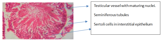
Figure 1. Testes of Rat (Control). A well-formed testicular vessel consisting of numerous seminiferous tubules embedded in matured interstitial tissues. The tissue is well lined by columnar epithelium of the Sertoli cells consisting of almost visible germ cells lined up in the tubular epithelium. The interstitial tissues consist of matured clusters of Leydig cells (H&E x 100).
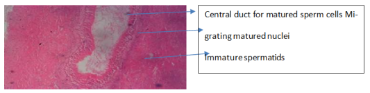
Figure 2. Epididymis of Rat (Control): Showing full single epididymis with a central duct canal and a lumen with clusters of spermatozoa. The small dense head tapers into the vas deferens (H&E x 100).
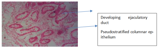
Figure 3. Vas deferens of Rat (Control): Showing forming ejaculatory ducts lined by lamina propria terminating in the pseudostratified columnar epithelium (H&E x 100).
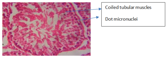
Figure 4. Seminal vesicle of Rat (Control): Showing well formed with coiled tubular muscle walls. Maturing sperm cells appear with dot micronuclei (H&E x 100).
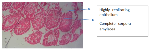
Figure 5. Prostate of Rat (Control): Showing well-formed highly replicating prostate epithelium that is highly lined columnar and secretory cells. Also, glandular lumen contains adequate corpora amylacea (H&E x 100).
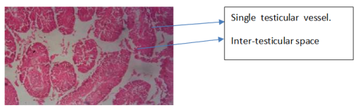
Figure 6. Testes of Rat given 2mg/kg of cadmium chloride. It consists of several columnar epithelium separated from each other and they are less visible in maturity and function. Sertoli are poorly lined and interspersed in the individual rete testis. (H&E x 100).
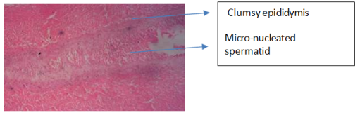
Figure 7. Epididymis of Rat given 2mg/kg of cadmium chloride. It shows clumsy head with vacuolated tail surrounded by unstriated smooth muscles (H&E x 100).

Figure 8. Testes of Rat given 4mg/kg of cadmium chloride. Showing several columnar epithelia that are widely separated from each other and they are less visible in maturity and function.
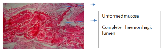
Figure 9. Vas deferens of Rat given 4mg/kg of cadmium chloride. showing haemorrhagic lamina propria. The entire lumen mucosa is unformed (H&E x 100).
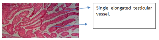
Figure 10. Testes of Rat given 6mg/kg of cadmium chloride. Rete testis is becoming smaller and elongated with few haemorrhages at the central spot. Epididymal interstitial ducts are poorly formed (H&E x 100).
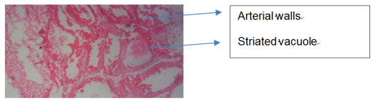
Figure 11. Rat seminal vesicle of Rat given 6mg/kg of cadmium chloride. Seminal vesicle battles proper formation of muscular walls with partial necrotic wall populated with arterial supply (H&E x 100).
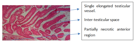
Figure 12. Testes of Rat given 8mg/kg of cadmium chloride. Rete testis is becoming smaller and elongated with few haemorrhages and partially necrotic at the anterior and posterior end.
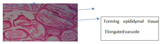
Figure 13. Epididymis of Rat given 500mg/kg of omega 3 fatty acid. Epididymis is well-formed with no negative pathological features detected. Each consist of nearly visible epididymal duct, a head, a tail and well-formed epididymal lumen (H&E x 100).
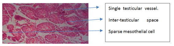
Figure 14. Testes of Rat given 1000mg/kg of omega- 3 fatty acid. Showing several well-formed testicular vessels consisting of maturing seminiferous tubules embedded in interstitial tissues, but all having fatty fibromuscular stroma. The tissues of individual rete testis are sparsely lined by columnar epithelium of the Sertoli cells (H&E x 100).
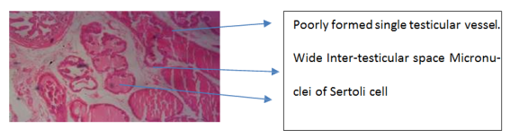
Figure 15. Testes of Rat given 2mg/kg cadmium chloride + 500mg/kg of omega-3 fatty acid. Several un-formed testicular vessels are seen with little fatty fibromuscular stroma. The peritoneal serosa tissues of rete testis are sparsely lined by mesothelial cells (H&E x 100).
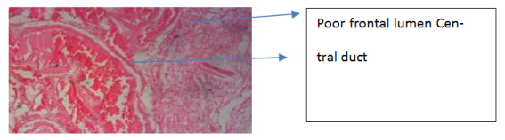
Figure 16. Epididymis of Rat given 2mg/kg cadmium chloride + 500mg/kg of omega-3 fatty acid. It shows poorly formed posterior epididymal lumen, while the anterior lumen is formed but has haemorrhagic appearance around the central duct (H&E x 100).
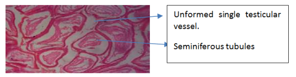
Figure 17. Testes of Rat given 4mg/kg cadmium chloride + 500mg/kg of omega-3 fatty acid. It shows a slight washing away of the thickened surface of the tunica albuginea of the entire rete testis leading to poor formation of the tunica vaginalis and visceral peritoneum (H&E x 100).
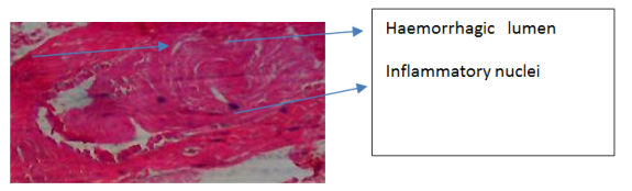
Figure 18. Vas deferens of Rat given 4mg/kg cadmium chloride + 500mg/kg of omega-3 fatty acid. It shows complete haemorrhagic lumen and lamina propria is poorly formed (H&E x 100).
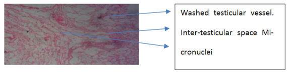
Figure 19. Testes of Rat given 6mg/kg cadmium chloride + 500mg/kg of omega-3 fatty acid. It shows complete washing away of the thickened surface of the tunica albuginea of the entire rete testis leading to poor formation of the fibrous septa with complete haemorrhagic tissue of the entire lumen having specific micronucleus of the stereocilia (H&E x 100).
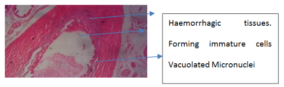
Figure 20. Seminal vesicle of Rat given 6mg/kg cadmium chloride + 500mg/kg of omega-3 fatty acid. Seminal vesicle wall is haemorrhagic with necrotic displasia and centrally vacuolated with arterial supply (H&E x 100).
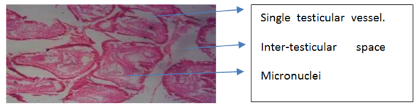
Figure 21. Testes of Rat given 8mg/kg cadmium chloride + 500mg/kg of omega-3 fatty acid. Each peritoneal cavity shows a very poor formation of mesothelial cells lining the tunica of the seminiferous tubules and epididymis.
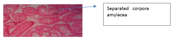
Figure 22. Prostate of Rat given 8mg/kg cadmium + 500mg/kg of omega-3 fatty acid. It shows a highly haemorrhagic replicating prostate epithelium that is lined with few columnar and secretory cells. The glandular lumen has adequate corpora amylacea separations (H&E x 100).
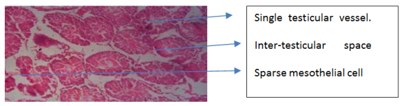
Figure 23. Testes of Rat given 2mg/kg cadmium chloride + 1000mg/kg of omega-3 fatty acid. Several well-formed testicular vessels consisting of maturing seminiferous tubules embedded in interstitial tissues, but all having fatty fibromuscular stroma. The tissues of individual rete testis are sparsely lined by columnar epithelium of the Sertoli cells (H&E x 100).
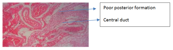
Figure 24. Epididymis given 2mg/kg cadmium chloride + 1000mg/Kg of omega-3 fatty acid. It shows formed and forming epididymal tissues consisting of ducts and seminiferous tubules of the apical stereocilia (H&E x 40).
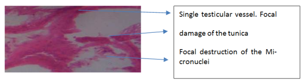
Figure 25. Testes of Rat given 4mg/kg cadmium chloride + 1000mg/Kg of omega-3 fatty acid. Photomicrograph shows complete focal destruction of rete testis, tunica vaginalis and tunica albuginea leading to very poor mesothelial cells of the serosa (H&E x 100).
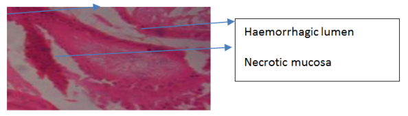
Figure 26. Vas deferens given 4mg/kg cadmium chloride + 1000mg/Kg of omega-3 fatty acid. It shows entirely diffused interstial haemorrhagic lumen mucosa and dysplasia (H&E x 100).
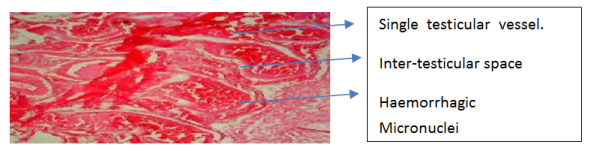
Figure 27. Testes of Rat given 6mg/kg cadmium chloride + 1000mg/Kg of omega-3 fatty acid. It shows complete focal destruction of rete testis, tunica vaginalis and tunica albuginea leading to central haemorrhage of the mesothelial cells of the serosa (H&E x 100).
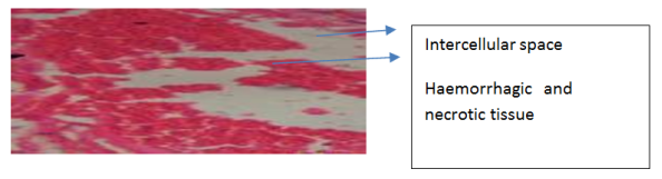
Figure 28. Seminal vesicle of Rat given 6mg/kg cadmium chloride + 1000mg/Kg of omega-3 fatty acid. It shows completely damaged with no proper formation of muscular walls and having partial necrotic wall populated with arterial supply (H&E x 40).
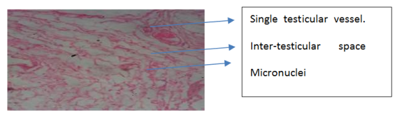
Figure 29. Testes given 8mg/kg cadmium chloride + 1000mg/kg of omega-3 fatty acid. It shows complete washing away of the thickened surface of the tunica albuginea of the entire rete testis leading to poor formation of the fibrous septa (H&E x 40).
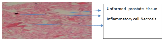
Figure 30. Prostate of Rat given 8mg/kg cadmium chloride + 1000mg/Kg of omega-3 fatty acid. It shows completely unformed with necrosis. There is no distinct prostate epithelium and glandular lumen has no adequate corpora amylacea separations (H&E x 100).